Figure, B-Mode ultrasound showing main portal] - StatPearls
4.9 (679) In stock
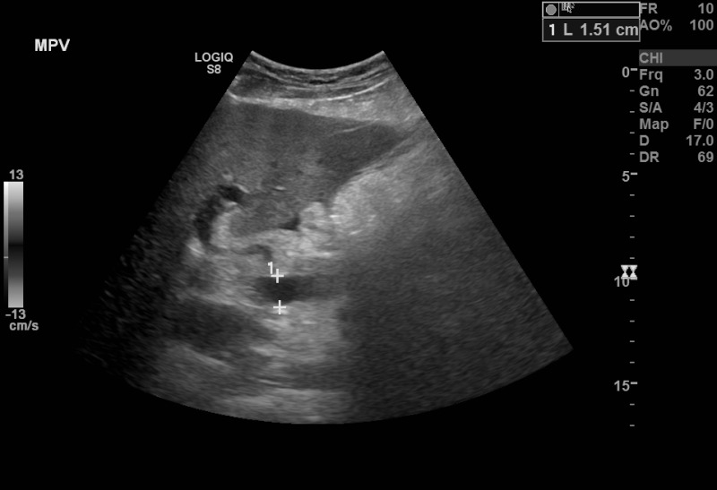
B-Mode ultrasound showing main portal vein diameter of 15.1 millimeters. This is an indirect finding of portal hypertension. Contributed by Brian Covello, MD
B mode ultrasound image (A) shows the hypoechoic tract (arrow) which is

Quantitative ultrasonographic examination of cerebral white matter by pixel brightness intensity as marker of middle-term neurodevelopment: a prospective observational study
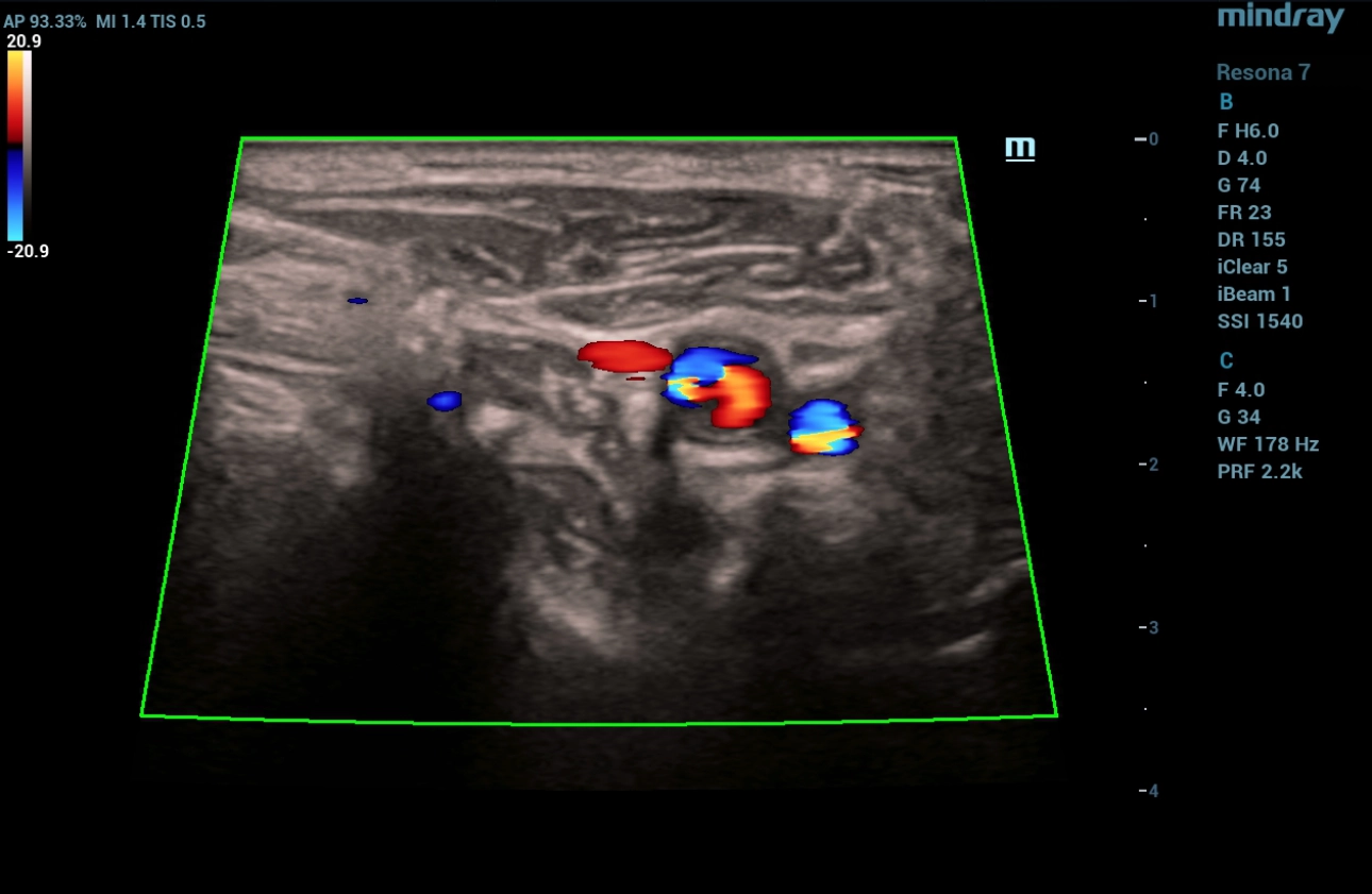
Ultrasound Journal 23 - Postoperative Ultrasound: A Case Study in Cardiovascular Pathology - Mindray
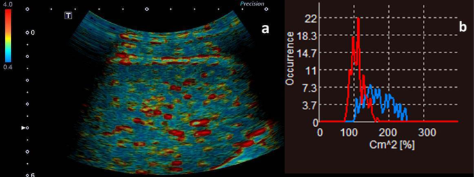
The severity of portal hypertension by a non-invasive assessment: acoustic structure quantification analysis of liver parenchyma, BMC Medical Imaging

Rad Tech CE, ASRT, ARRT® CE, Category A Credits

Pathogens, Free Full-Text

Prospective Assessment of Treatment-Induced Liver Injury as a Cause of Diffuse Pathologic Hepatic Enhancement in Contrast-Enhanced Ultrasound - ScienceDirect

Assessment of the portal vein anatomy with 3 D ultrasound

Diagnostics, Free Full-Text
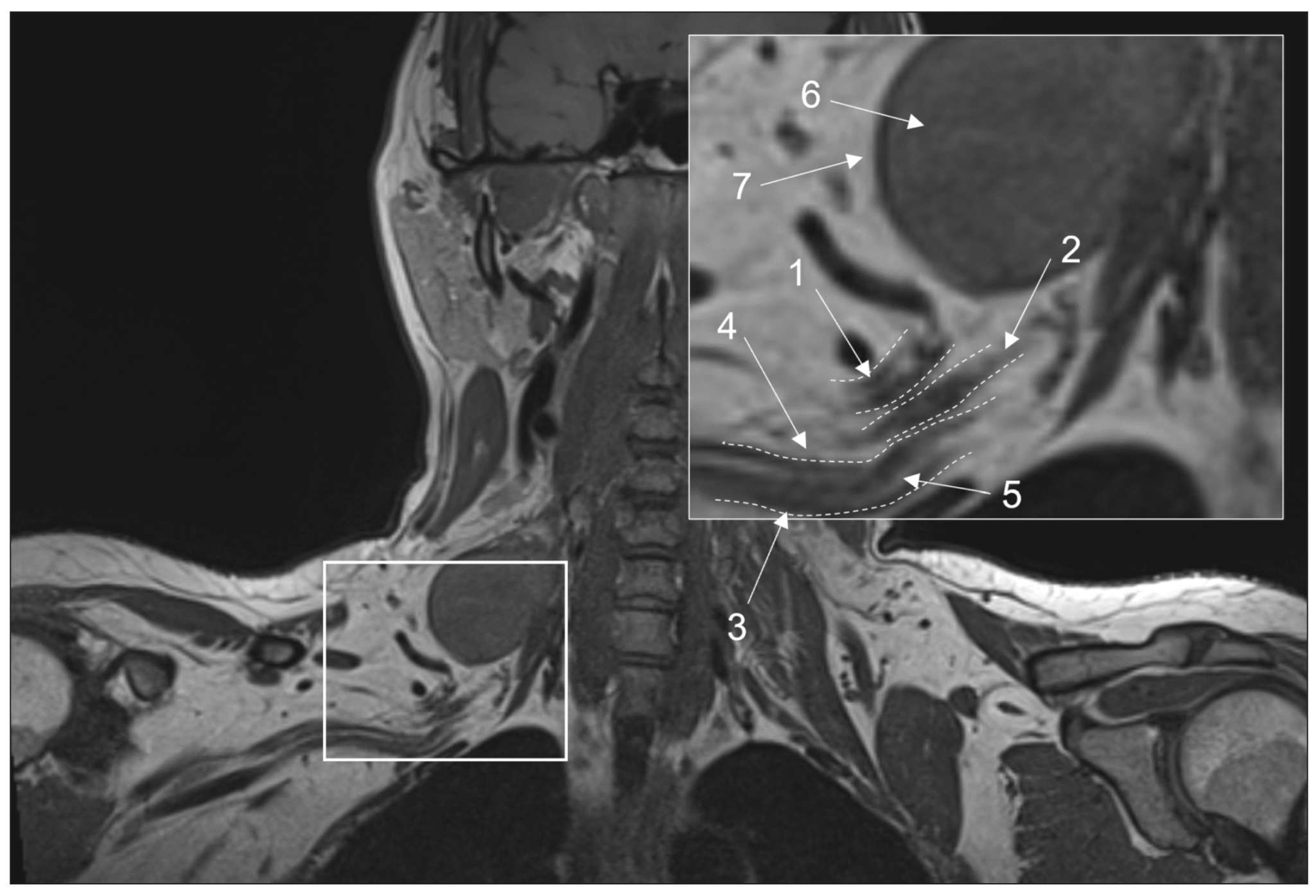
Conventional magnetic resonance imaging of peripheral nerves: MR-neurography - Morozova - Digital Diagnostics
B-mode of the ultrasound Download Scientific Diagram
Physics and Instrumentation in Doppler and B-mode Ultrasonography
 Glamorise Women's Plus Size MagicLift Seamless Sports Bra Wirefree 1006
 Women's Satin Silky Dress Casual Elastic High Waist Stretch Elegant Pants Trousers
 VICTORIA'S SECRET VERY SEXY Black Lace Mesh Cheeky Panty S M L Cutout Open Back
- Downloadable Herobrine Minecraft Skin, HD Png Download , Transparent Png Image - PNGitem, herobrine skin png
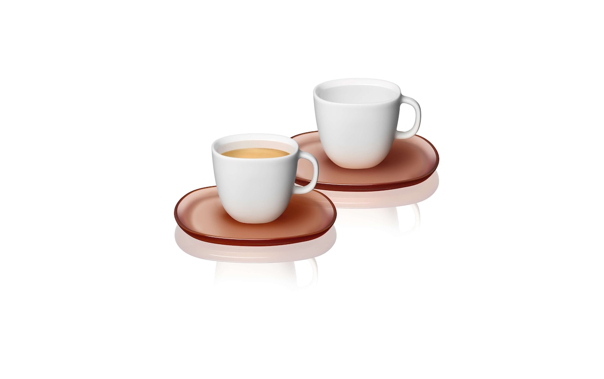 LUME Espresso Cups
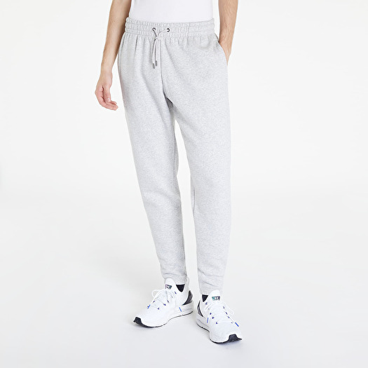 Pants and jeans Under Armour Essential Fleece Jogger Ghost Gray Medium Heather/ White
