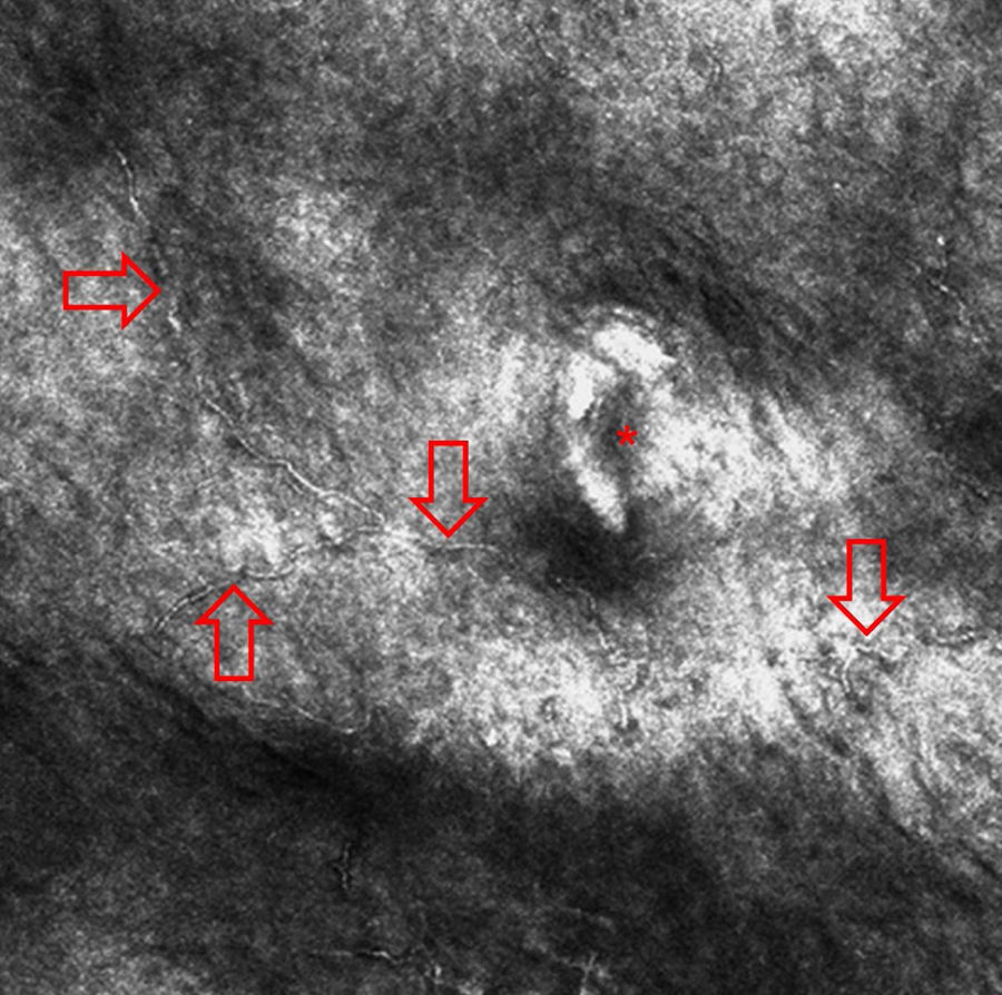
Beware of reflectance confocal microscopy artifacts when searching
4.8 (298) In stock

Clinical appearance of tinea nigra (highlighted by the circle)

Jean PERROT, Medical Doctor, Professor, Centre Hospitalier Universitaire de Saint-Étienne, Saint-Étienne, CHU St Etienne, Department of Dermatology

PDF) Cell-Specific Markers for the Identification of Retinal Cells by Immunofluorescence Microscopy

Reflectance confocal microscopy for cutaneous infections and infestations

SciELO - Brasil - Beware of reflectance confocal microscopy artifacts when searching hyphae in acral skin - Reply, Beware of reflectance confocal microscopy artifacts when searching hyphae in acral skin - Reply,

White piedra, black piedra, tinea versicolor, and tinea nigra: contribution to the diagnosis of superficial mycosis. - Abstract - Europe PMC

Grain density (~o) over fungal structures in relation to the grain
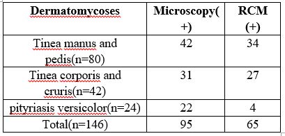
The challenge of Diagnosing Common Dermatomycosis by Reflectance Confocal Microscopy

Focal Volume Optics and Experimental Artifacts in Confocal Fluorescence Correlation Spectroscopy - ScienceDirect

Morphological and anatomical features of the basal and medial zones of

Learn To Minimize Artifacts In Fluorescence Microscopy
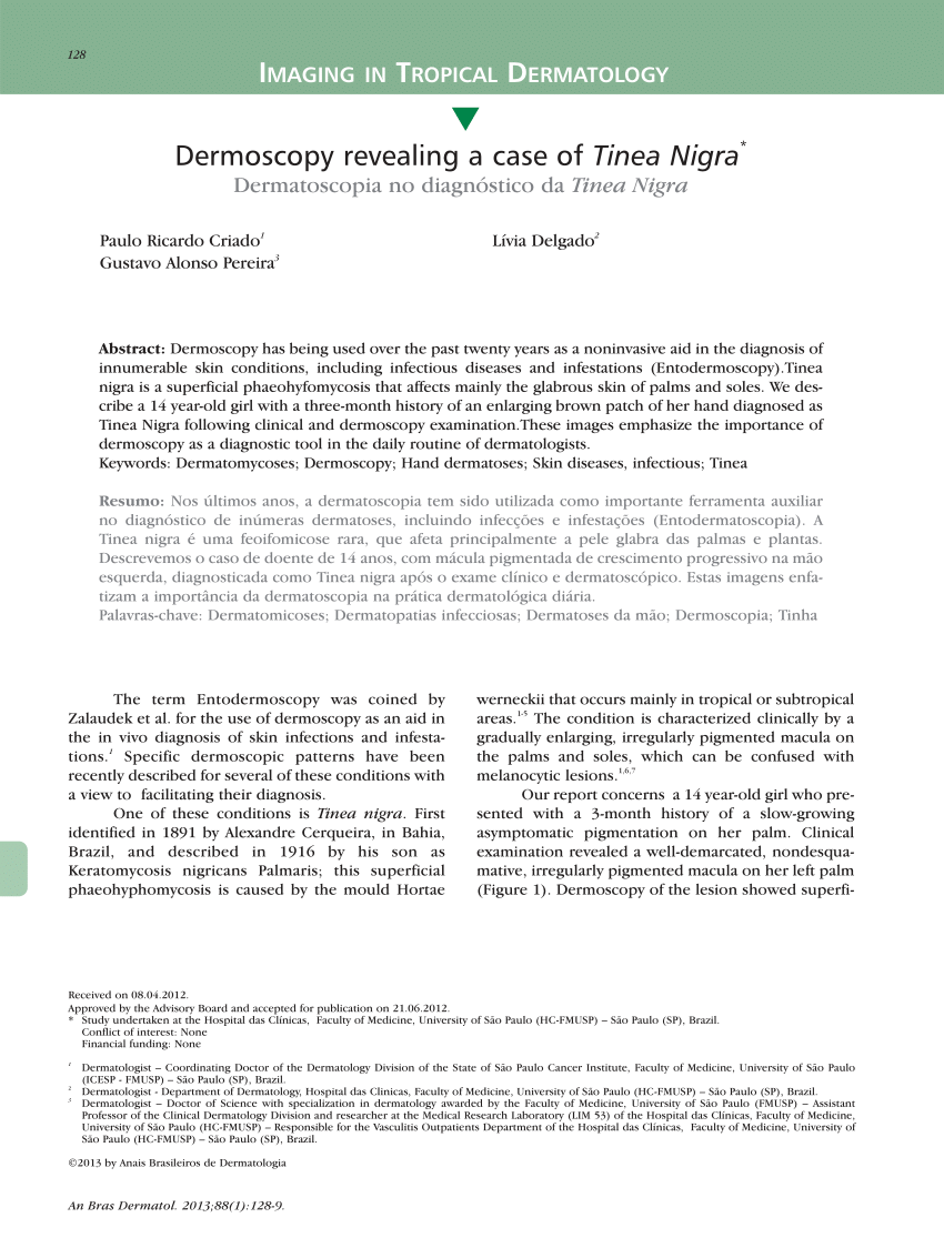
PDF) Dermoscopy revealing a case of Tinea Nigra

Dermoscopy revealing a case of Tinea Nigra. - Abstract - Europe PMC

Assays for the localization of green fluorescent protein (GFP)-FgAtg8
Fungal infections By: amin alajlouni. - ppt download
Dermoscopy Atlas Diagnosis Detail
This is a photomicrograph of the fungus Hortaea werneckii, the
 Zara Wide Leg Brown Leather Pants
- Zendaya Wore a Bra Top With Star-Shaped Boob Cutouts to the NAACP
 Fila Activewear for Men, Online Sale up to 81% off
 Urbanic - Moda de Londres. Nós somos porque você é!
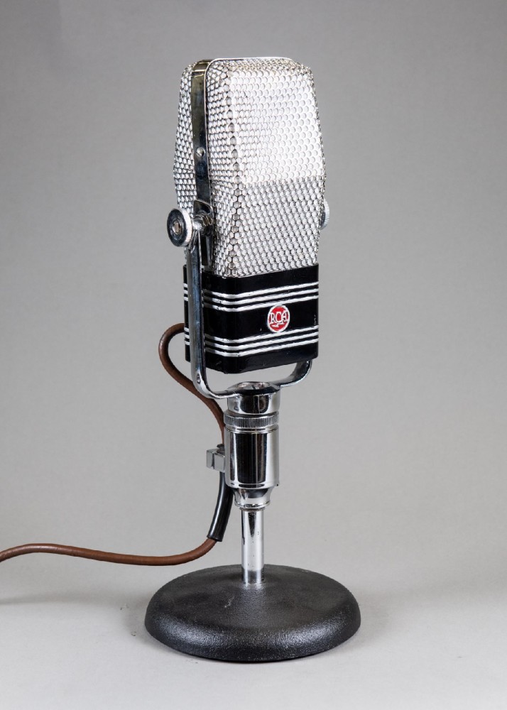 Microphone, RCA 44B, With Yoke Mount And Cable, Non-Operational, Black, RCA, 1930s+, Metal, 12H, 5W, 3L - History For Hire
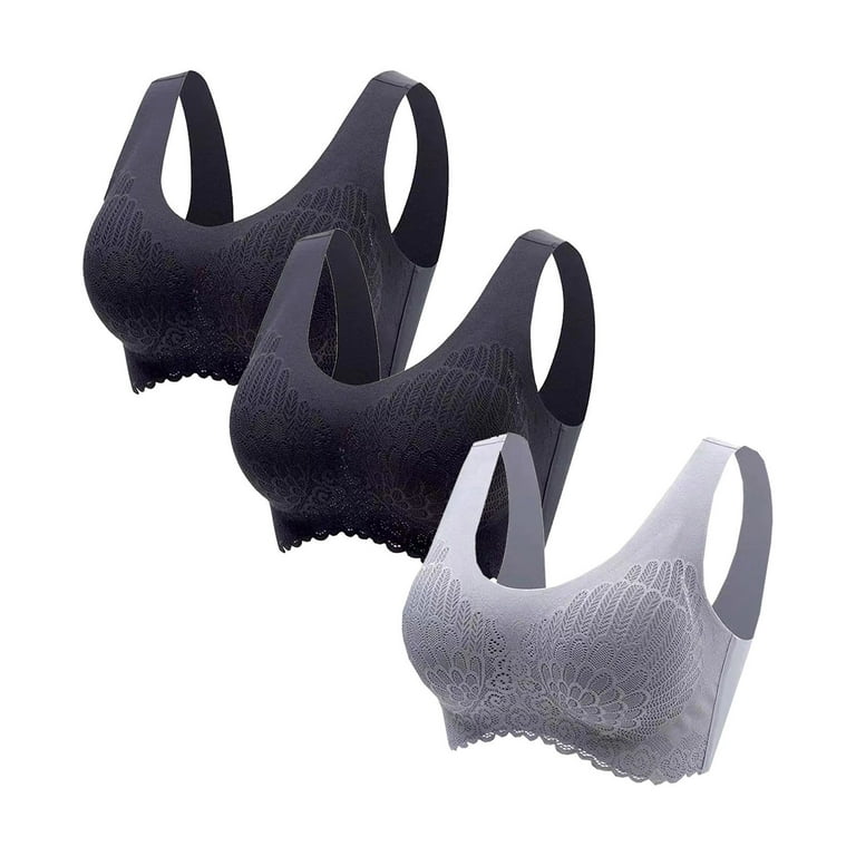 Mrat Clearance Bras for Women Clearance Women Tops Bra Wire-Free