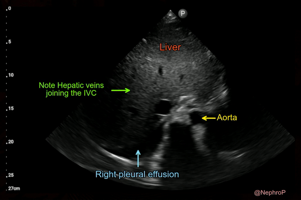
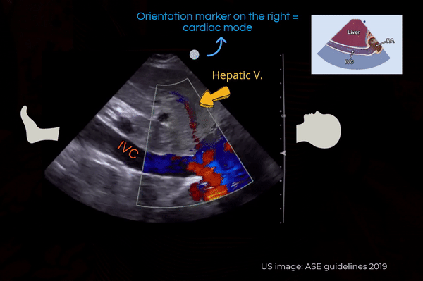
Pitfalls of inferior vena cava M-mode – NephroPOCUS
4.9 (321) In stock

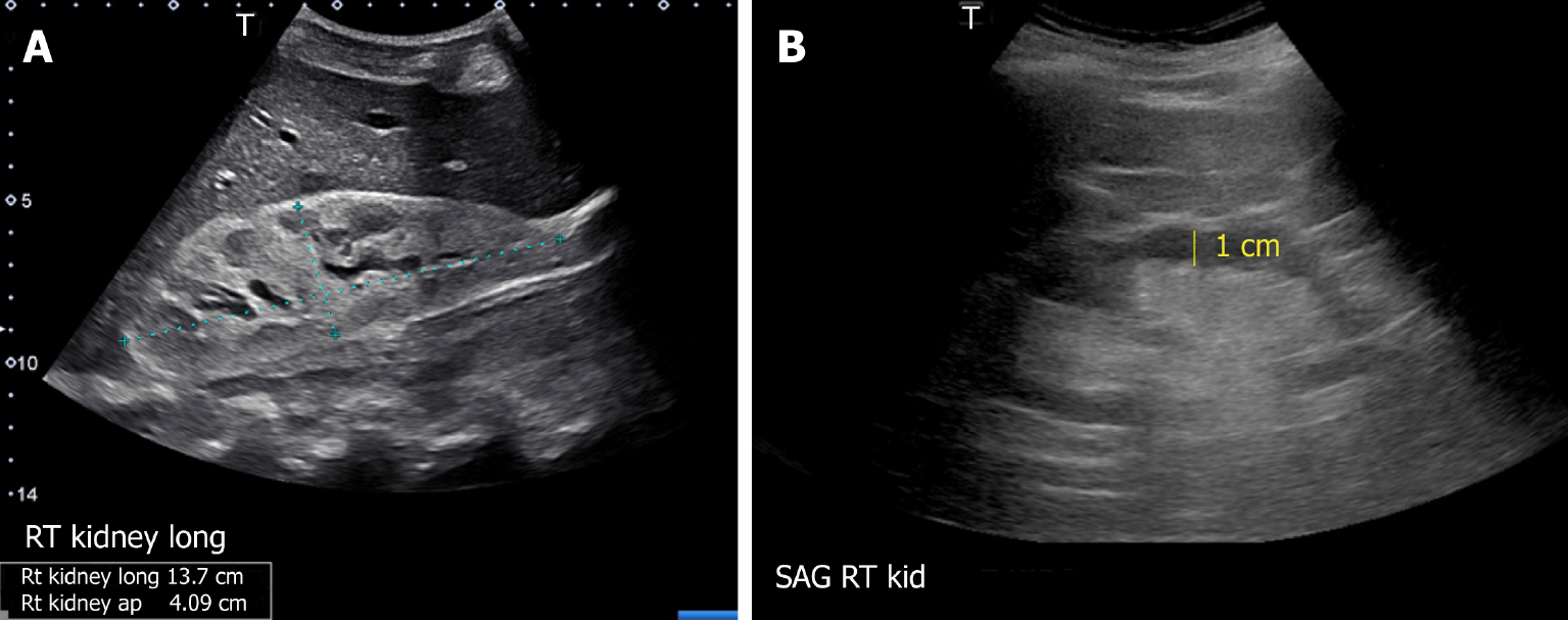
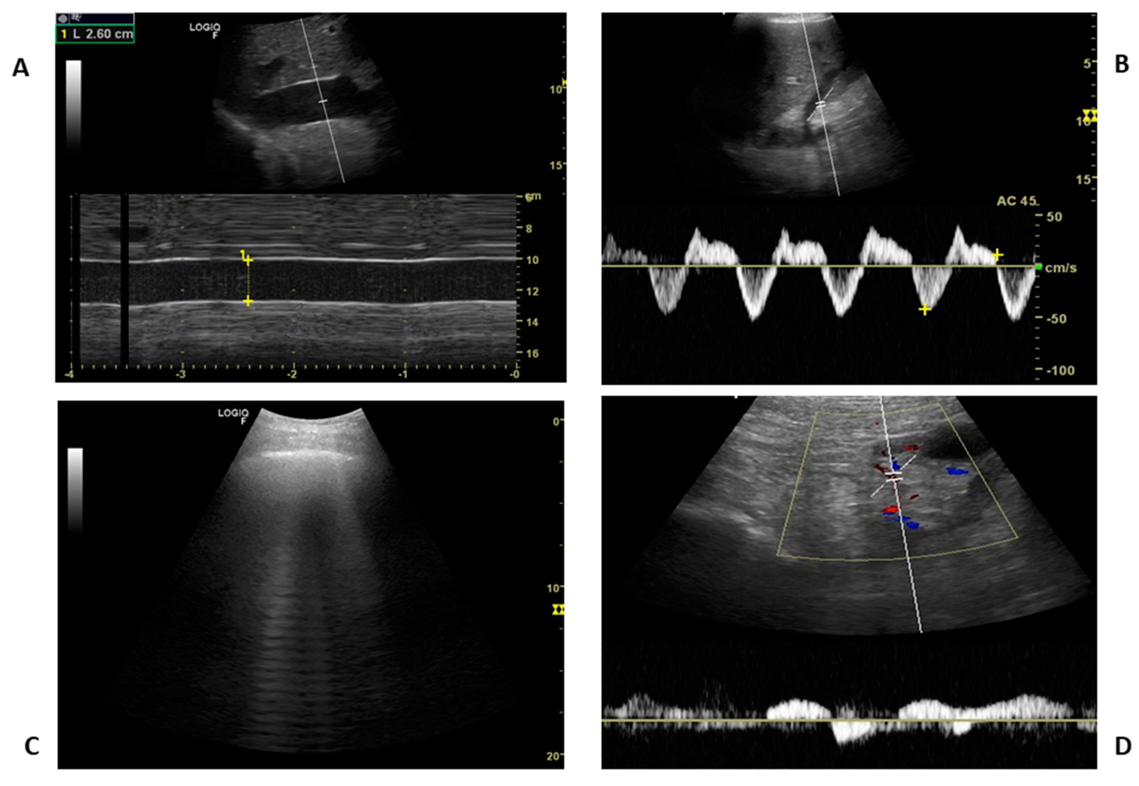
Visual estimation of IVC collapse on B-mode (grey scale image) is generally preferred to M-mode, though in theory, M-mode measurement might be able to give accurate collapsibility index. There are several reasons for this. A major limitation of IVC M-mode is that the vessel moves mediolaterally and craniocaudally during respiration, with collapse occurring off axis…

PoCUS in nephrology: a new tool to improve our diagnostic skills
Transcending boundaries: Unleashing the potential of multi-organ
JCM, Free Full-Text

Image Acquisition Method for the Sonographic Assessment of the
Thread by @NephroP on Thread Reader App – Thread Reader App

PoCUS in nephrology: a new tool to improve our diagnostic skills
Thread by @NephroP on Thread Reader App – Thread Reader App
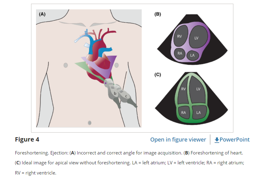
Links To And Excerpts From The Second E - Ejection From The 5Es

Abhilash Koratala – Page 9 – NephroPOCUS
Inferior Vena Cava POCUS: The Basics of Image Acquisition - Renal

PDF) PoCUS in Nephrology: A new tool to improve our diagnostic skills

Venous Excess Doppler Ultrasound for the Nephrologist: Pearls and Pitfalls - ScienceDirect

PDF) Venous Excess Doppler Ultrasound for the Nephrologist: Pearls
Inferior Vena Cava POCUS: The Basics of Image Acquisition - Renal

Cardiac – Page 2 – NephroPOCUS
Echocardiography Tutorial - Echocardiographic Modes
Physics of Ultrasound - NYSORA
Modes Ultrasound A-mode- amplitude mode. B-mode- brightness mode
Ultrasound and non-ultrasound imaging techniques in the assessment
Internal carotid artery chronic occlusion: B-mode and colour
 Bandeau Bikini Tops USA Girls Shop Kulani Kinis Bikini Tops
 PERFUME FEMININO PRADA INFUSION D'IRIS EAU DE PARFUM_8435137710195
 Peach Blossom Print, Flower Art, Botanical Illustration, Wall Art
 93 Best One-Pot Meals, One-Pot Recipes & Ideas
- High-Rise Loose Flare Overalls in Demott Wash
 ALIX NYC - Pearson Ribbed-jersey Midi Dress - Black
